FIGURE 1. Morphology of cephalothorax and a general habitus of studied taxa. A, Cephalothorax of nephropid lobsters with selected morphological characters (modified from Tshudy and Sorhannus, 2003). B, Habitus of thaumastocheliform lobsters, exemplified by Thaumastocheles zaleucus (from Calman, 1911). C, Habitus of ctenochelid ghost shrimps, exemplified by Ctenocheles balssi (from Kishinouye, 1926). D, Groove pattern of Oncopareia bredai Bosquet, 1854 (redrawn from Tshudy and Sorhannus, 2000b, fig. 2.2). E, Groove pattern of Hoploparia beyrichi Schlüter, 1862 (redrawn from Tshudy and Sorhannus, 2000b, fig. 2.3).
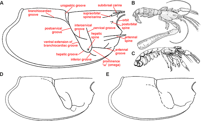
FIGURE 2. Oncopareia bredai (sensu Bosquet, 1854), as a composite species. A, Cephalothorax of O. bredai (from Bosquet, 1854, pl. 10, fig. 5). B, Pleon of Hoploparia beyrichi Schlüter, 1862 (from Bosquet, 1854, pl. 10, fig. 6). C, Claw of H. beyrichi (from Bosquet, 1854, pl. 10, fig. 71b). D, Composite drawing from Mertin (1941, text-fig. 9e). E, Pleonal segments 1 and 2 of O. bredai (as Homarus bosqueti Pelseneer, 1886; from Pelseneer, 1886, fig. 4).
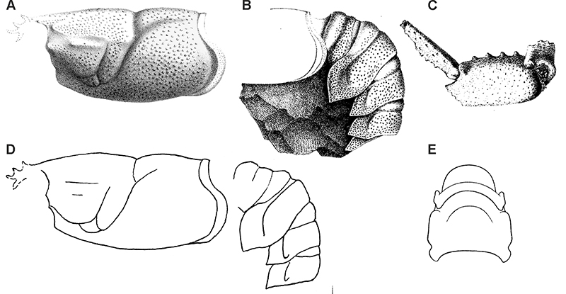
FIGURE 3. Oncopareia bredai Bosquet, 1854; upper Maastrichtian (Kunrade Formation) of the Kunrade area, southern Limburg, the Netherlands. A, Cephalothorax in lateral view (from Bosquet, 1854, pl. 10, fig. 5). B, Anterior portion of cephalothorax in lateral view (from Bosquet, 1854, pl. 10, fig. 8). C, Cephalothorax with pleon and tailfan in lateral view (from Bosquet, 1854, pl. 10, fig. 61). D, Cephalothorax in lateral view (IRScNB 90-32c). E, Cephalothorax in lateral view (IRScNB 90-17). F, Anterior portion of cephalothorax in lateral view (IRScNB 90-15b). G, Incomplete individual in lateral view (IRScNB 90-33f, designated lectotype herein). H, I, Pleon in lateral (H) and dorsal (I) views (IRScNB 90-35c). J, Pleonal segments 1-3 in lateral view (IRScNB 90-23b). K, Right (crusher) and left (pectinate) claws of the same individual (IRScNB 90-28). L, Fingers of pectinate claw in lateral view (IRScNB 90-3b). M, Crusher claw in lateral view (IRScNB 90-32a). N, Pectinate claw in lateral view (IRScNB 90-27b). Specimens in D-F and H-N were whitened with ammonium chloride prior to photography.
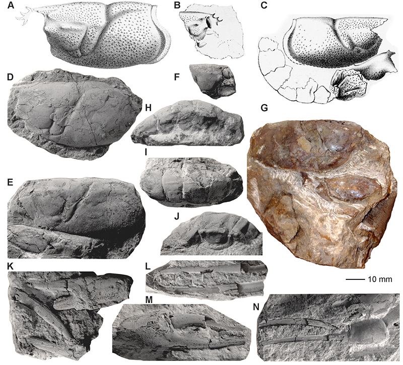
FIGURE 4. Oncopareia bredai Bosquet, 1854; cephalothorax with associated pleonal segments 1 and 2 and both claws (MNHN R03408) from the upper Maastrichtian (Kunrade Formation) at Kunrade, the Netherlands. A, Cephalothorax in left lateral view. B, Cephalothorax in dorsal view. C, Cephalothorax in right lateral view. D, Both claws. E, Crusher claw in lateral view.
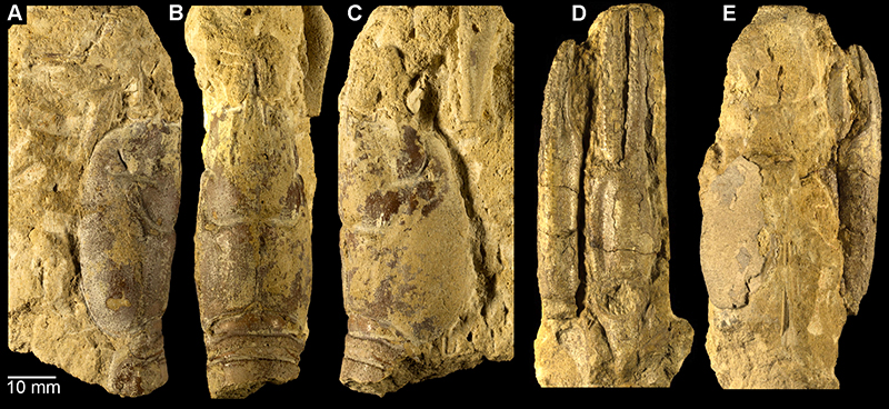
FIGURE 5. Oncopareia bredai Bosquet, 1854, cephalothoraxes, partial pleons and isolated claws; with exception of H (which is from the upper Campanian Zeven Wegen Member), all are from the Vijlen Member, interval 6 (upper lower Maastrichtian) at the CPL/CBR quarry complex near Haccourt and Lixhe, north-east Belgium. A, Cephalothorax in right lateral view (NHMM JJ 15836). B, C, Cephalothorax in right lateral view, and counterpart (NHMM JJ 6741). D, E, Cephalothorax in right lateral view, and counterpart (NHMM JJ 14618). F, Fragmentary cephalothorax and pleon in right lateral view (NHMM JJ 6764). G, Isolated pleura (NHMM JJ 6741). I, Fragment of cephalothorax and pectinate claw (NHMM JJ 14618). J, pectinate claw in lateral view (NHMM JJ 15534). K, crusher claw in lateral view (NHMM JJ 7194). L, fragmentary pectinate claw in lateral view (NHMM JJ 6839).
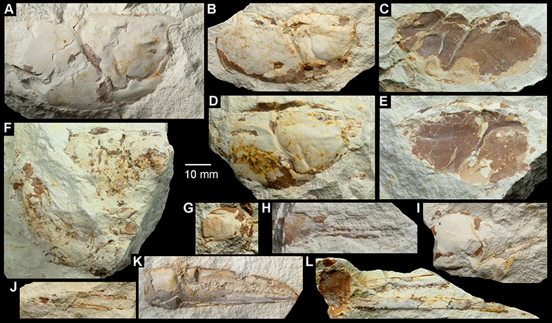
FIGURE 6. Oncopareia bredai Bosquet, 1854; isolated cephalothoraxes from the upper Turonian (uppermost part of the Jizera Formation) at Vinary near Vysoké Mýto, Czech Republic. A, Isolated cephalothorax in lateral view (NM O3470). B, Digital copy of Fritsch and Kafka (1887, pl. 5, fig. 2). C, Isolated cephalothorax in lateral view (NM O6861). Both specimens were whitened with ammonium chloride prior to photography.
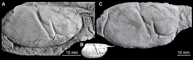
FIGURE 7. Nymphaeops coesfeldiensis Schlüter, 1862 (= Oncopareia bredai Bosquet, 1854, as here understood). A, Incomplete cephalothorax with associated pleon in lateral view (from Schlüter, 1862, pl. 13, fig. 3). B, Cephalothorax and pleon in dorsal view (from Schlüter, 1862, pl. 13, fig. 6). C, Reconstruction of cephalothorax in dorsal view (from Schlüter, 1879, pl. 15, fig. 2). D, Near-complete individual in lateral view (from Schlüter, 1879, pl. 15, fig. 1). E, Cephalothorax and pleon in lateral view (from Mertin, 1941, text-fig. 9a). F, Pectinate claw (from Mertin, 1941, text-fig. 10k). G, Crusher claw (from Mertin, 1941, text-fig. 10b). H, Crusher claw (from Mertin, 1941, text-fig. 10d). Nymphaeops coesfeldiensis Schlüter, 1862 (= Oncopareia bredai Bosquet, 1854, as here understood) from the Vaals Formation (lower Campanian) at the former CPL SA quarry, Haccourt, north-east Belgium (I-K). I, Near-complete cephalothorax with associated appendages in lateral view (NHMM JJ 9974). J, Crusher claw (NHMM HB 393). K, Fingers of pectinate claw (NHMM HB 756). Figure parts A-H not to scale.
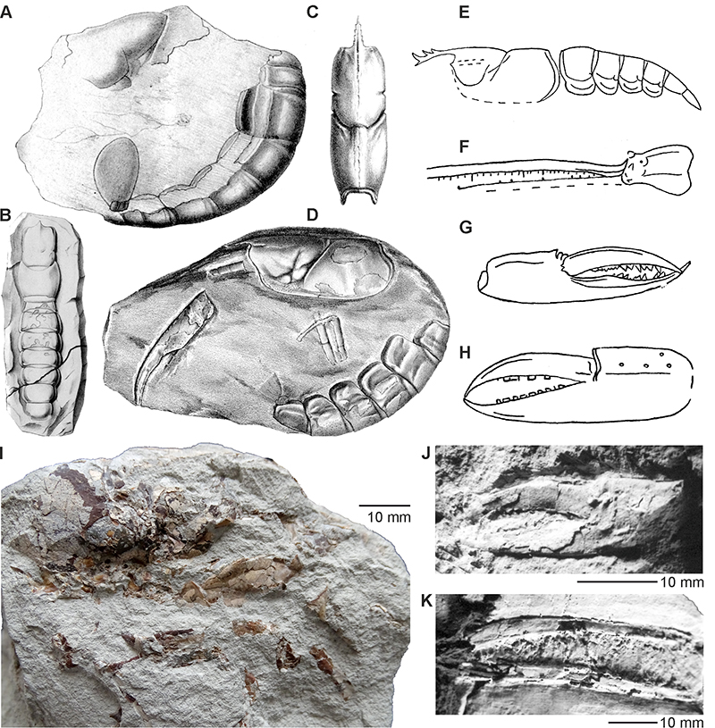
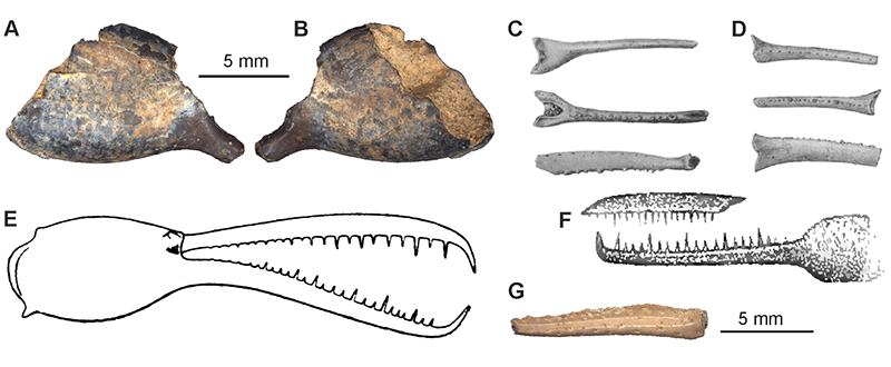
FIGURE 8. Species of Oncopareia based on fragmentary specimens. A-E. Oncopareia esocina (Fritsch, in Fritsch and Kafka, 1887); pectinate claw with associated pleon and tailfan from the lower Coniacian (Teplice Formation) at Oškobrh near Poděbrady, Czech Republic. A, Digital copy of figure from Fritsch and Kafka (1887, text-fig. 59). B, Digital copy of figure from Fritsch and Kafka (1887, pl. 4, fig. 7). C, Part of the holotype (NM O3467). D, Counterpart of the holotype (NM O3468). E, Detailed view of pectinate claw. F, G, Oncopareia klintebjergensis Jakobsen and Collins, 1979; Paleocene (Lellinge Greensand Limestone) of Klintebjerg, Denmark. F, Incomplete cephalothorax in lateral view (holotype). G, Fingers of pectinate claw (paratype).
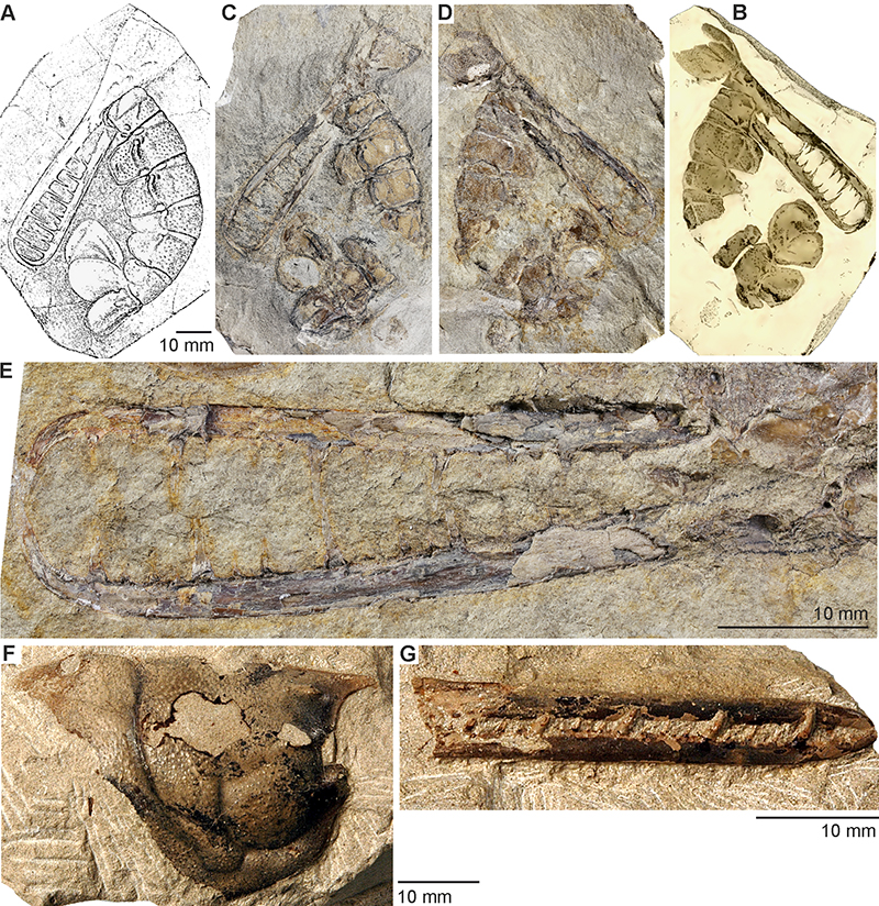
FIGURE 9. Oncopareia lunata (Fritsch, in Fritsch and Kafka, 1887); incomplete cephalothorax with pleon from the lower–middle Turonian (Bílá Hora Formation) in Prague, Czech Republic (NM O3473). A, Digital copy of the figure from Fritsch and Kafka (1887, pl. 5, fig. 6). B, Holotype in dorsal view with morphological features highlighted by pencil (presumably by Fritsch himself). C, Holotype in dorsal view with pencil marks removed. D, Holotype in lateral view.
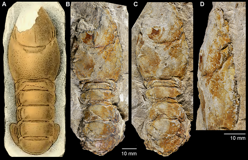
FIGURE 10. Hoploparia beyrichi Schlüter, 1862; upper Maastrichtian (Kunrade Formation), Kunrade area, southern Limburg, the Netherlands. A, Pleon in lateral view (from Bosquet, 1854, pl. 10, fig. 6). B, Right claw in inner and outer lateral views (from Bosquet, 1854, pl. 10, fig. 71). C, Incomplete right claw (from Bosquet, 1854, pl. 10, fig. 7). D–E, Pleon in dorsal (D) and lateral (E) views (IRScNB 90-32f). F–H, Fragmentary claw (right propodus) in inner lateral (F), outer lateral (G) and dorsal (H) views (IRScNB 90-19j). I, Cephalothorax with associated incomplete pleon in lateral view (IRScNB 90-32d). J, Right claw (propodus and carpus) in inner lateral view (IRScNB 90-24b). K, Cephalothorax with associated pleon (holotype, MB. A. 209). L–M, Cephalothorax in lateral (L) and dorsal (M) views (IRScNB 90-33g). Specimens were whitened with ammonium chloride prior to photography.
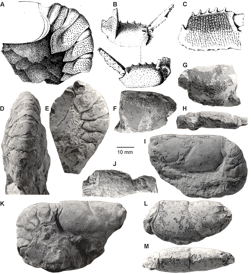
FIGURE 11. Hoploparia biserialis Fritsch, in Fritsch and Kafka, 1887; lower–middle Turonian (Bílá Hora Formation) in Prague, Czech Republic. A, Cephalothorax with pleon and tailfan in lateral view (lectotype, NM O4044). B-C, Isolated right cutter claw in dorsal (B) and lateral (C) views (NM O3457). Specimen in A was whitened with ammonium chloride prior to photography.
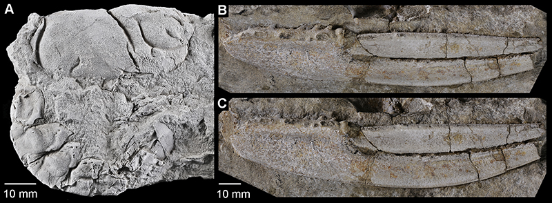
FIGURE 12. Astacidean lobsters with uncertain affinities. A-B, “Hoploparia” macrodactyla Schlüter, in von der Marck and Schlüter, 1868; fragmentary cephalothorax with associated claws (from Schlüter, 1862, pl. 11, fig. 5). C, “Nymphaeops” belgicus Forir, 1887; cephalothorax in dorsal view (from Forir, 1887, pl. 7, fig. 1). D, “Nymphaeops” sendenhorstensis Schlüter, 1862; cephalothorax with pleon and chelipeds (from Schlüter, 1862, pl. 14, fig. 5).
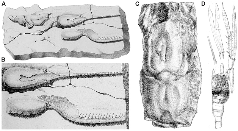
FIGURE 13. “Stenocheles” parvulus Fritsch, in Fritsch and Kafka, 1887; lectotype from the lower–middle Turonian (Bílá Hora Formation) in Prague, Czech Republic. A–C, Part of the specimen (NM O3455) under various light conditions. D–F, Counterpart of the same (NM O9092), including the historical cast (D).
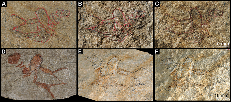
FIGURE 14. Non-astacidean decapod species with pectinate claws. A–B, Ctenocheles cookei (Rathbun, 1935), left propodus in inner (A) and outer (B) lateral views (holotype, USNM 371511). C–D, Ctenocheles cultellus (Rathbun, 1935), incomplete dactylus (A) and fixed finger (B) in upper, lower and lateral views (from Rathbun, 1935, pl. 14, figs. 7–12). E, Ctenocheles inaequidens (Pelseneer, 1886), right major claw (holotype, from Pelseneer, 1886, text-fig. 1). F, ? Ctenocheles pectiniformis (Böhm, 1891), isolated claw (from Böhm, 1891, pl. 1, fig. 2, 2a). G, “Ischnodactylus” dentatus Rathbun, 1935, isolated finger (USNM MO 495109).